
Emerging Mammography Trends Reshaping Radiology Operations in 2026
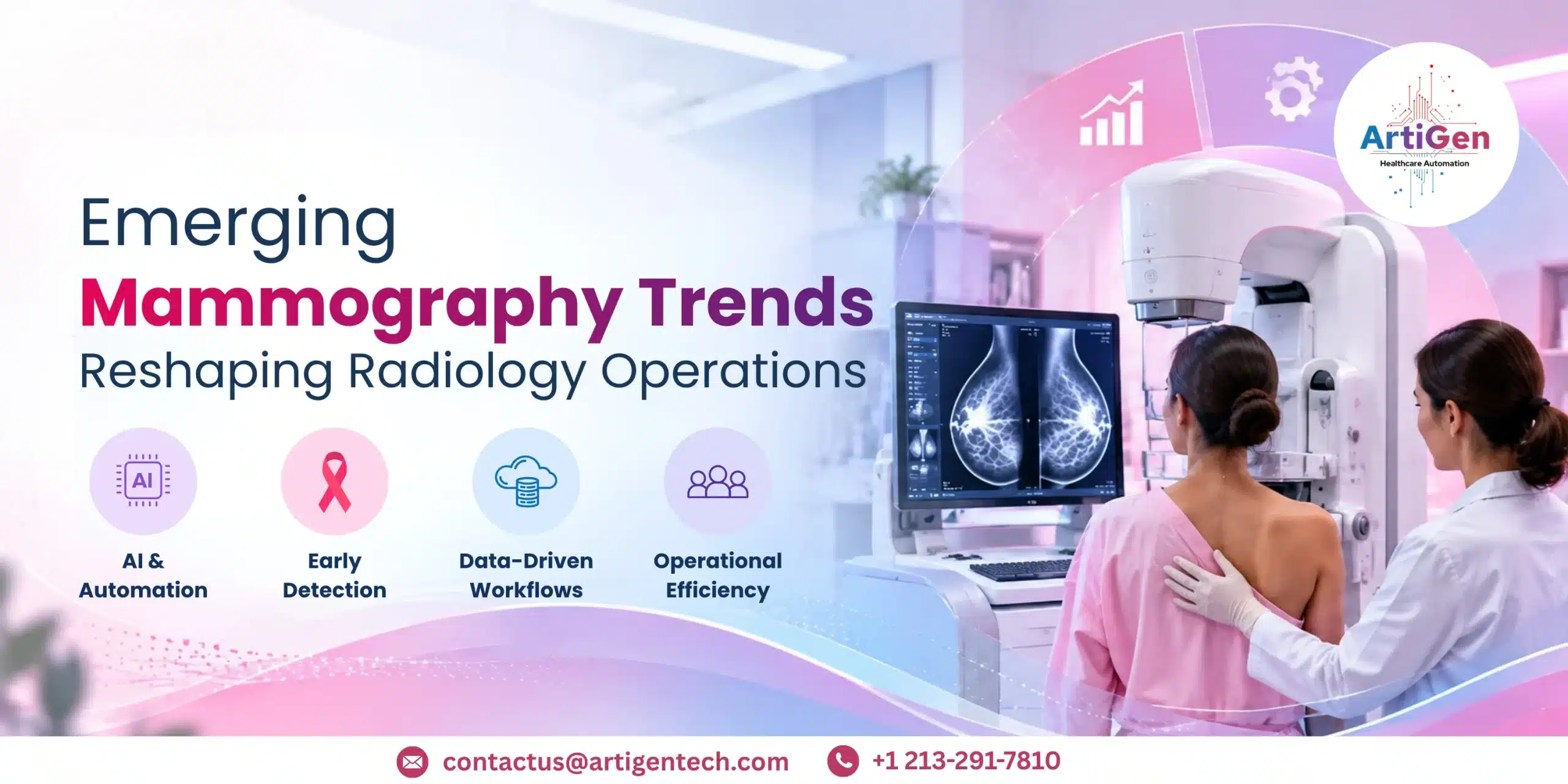
Mammography is moving beyond traditional screening models. In 2026, radiology organizations are managing increasing screening volumes, breast imaging staffing shortages, evolving breast cancer screening guidelines, growing awareness of dense breast tissue, and rapid advances in breast imaging technology. These changes are reshaping how healthcare organizations approach screening, diagnosis, staffing, and workflow management.
The combination of artificial intelligence, advanced imaging, risk-based protocols, and automation is creating a new model for breast imaging operations. Technologies such as digital breast tomosynthesis, contrast-enhanced mammography, and AI-assisted imaging are increasingly being incorporated into modern workflows. At the same time, mammography workflow automation and radiology workflow automation are helping organizations manage growing examination volumes while improving radiology operational efficiency.
AI Is Becoming Part of the Mammography Workflow
One of the most significant developments in modern breast imaging technology is artificial intelligence. AI is increasingly being used to support breast cancer detection, image analysis, case prioritization, and workflow management.
AI-assisted systems can identify suspicious imaging patterns and help prioritize examinations that may require additional review. This development in AI breast cancer detection can support radiologists managing high examination volumes while contributing to earlier identification of potentially concerning findings.
For organizations experiencing a breast imaging staffing shortage, AI can provide workflow support without replacing the clinical judgment of radiologists. When appropriately integrated, AI can reduce repetitive tasks, prioritize worklists, and support mammography workflow efficiency.
The important shift is that AI is moving from an isolated point solution toward an integrated part of breast imaging operations. Combining AI breast cancer detection with radiology workflow automation can help organizations create more connected imaging pathways.
Risk-Based Screening Is Reshaping Mammography
Traditional screening models are increasingly being complemented by individualized risk assessment. Risk-based breast cancer screening considers factors such as age, family history, breast density, previous findings, and other documented risk factors when determining appropriate screening pathways.
This makes breast cancer risk assessment an increasingly important component of modern breast imaging operations. Instead of treating every patient through exactly the same pathway, healthcare organizations can use documented risk information to determine appropriate screening and diagnostic follow-up.
A screening mammogram remains an important component of routine screening. However, the appropriate screening interval and additional imaging may depend on individual patient circumstances and applicable clinical recommendations.
Healthcare organizations therefore need to monitor current breast cancer screening guidelines, mammography screening guidelines, and breast imaging guidelines. These guidelines help organizations establish consistent processes for screening, follow-up, and diagnostic evaluation.
As risk-based breast cancer screening becomes more important, effective breast cancer risk assessment and guideline-driven workflows can help support breast cancer early detection.
Dense Breast Tissue Is Increasing Supplemental Imaging Needs
Another major consideration for breast imaging departments is dense breast tissue. Breast density can affect how abnormalities appear on mammography and may influence decisions regarding supplemental imaging based on individual risk and applicable guidelines.
A dense breast mammogram remains an important screening examination, but some patients with dense breast tissue may require additional imaging depending on their clinical circumstances.
This is increasing demand for coordinated diagnostic breast imaging. Organizations need efficient pathways for scheduling, performing, interpreting, and following up on additional examinations.
The growing focus on dense breast tissue is also influencing the adoption of advanced breast imaging technology. Radiology organizations need systems capable of handling screening mammography, supplemental imaging, reporting, and follow-up without creating unnecessary workflow delays.
For patients receiving a dense breast mammogram, the operational objective should be a coordinated pathway that connects screening findings with appropriate diagnostic evaluation.
Digital Breast Tomosynthesis Is Reshaping Imaging
Digital breast tomosynthesis, commonly called 3D mammography, is an important development in modern breast imaging. Instead of producing only conventional two-dimensional images, digital breast tomosynthesis acquires multiple low-dose projections that are reconstructed into a series of images.
The wider use of digital breast tomosynthesis is influencing equipment utilization, image interpretation, storage, reporting, and radiologist workload. As this technology becomes more integrated into routine services, organizations must consider how it fits into existing breast imaging operations.
Advanced breast imaging technology is valuable only when it is connected to efficient workflows. Scheduling, image transfer, PACS integration, reporting, quality assurance, and follow-up processes all need to work together.
For this reason, the adoption of digital breast tomosynthesis is not simply an equipment decision. It is part of a broader strategy for improving mammography workflow efficiency and radiology operational efficiency.
Contrast-Enhanced Mammography Is Gaining Attention
Contrast-enhanced mammography is another technology receiving increased attention in breast imaging. CEM combines mammographic imaging with intravenous contrast administration to provide additional information about areas of abnormal enhancement in selected clinical situations.
The role of contrast-enhanced mammography can include specific diagnostic evaluations and treatment-response assessment, depending on clinical requirements and institutional protocols.
As the use of contrast-enhanced mammography expands, imaging organizations need to consider patient preparation, contrast administration, scheduling, safety procedures, image interpretation, and coordination with other diagnostic services.
This makes CEM both a technology and an operational consideration. Effective breast imaging operations need workflows that can accommodate these additional requirements without creating unnecessary delays in diagnostic breast imaging.
Mammography Workflow Automation Is Becoming Essential
Growing examination volumes and limited specialist capacity are making mammography workflow automation increasingly important. Automation can support scheduling, worklist management, image routing, reporting processes, follow-up tasks, and other repetitive administrative activities.
The primary objective of mammography workflow automation is to reduce unnecessary manual intervention and improve mammography workflow efficiency.
At the enterprise level, radiology workflow automation can connect mammography with broader imaging and reporting systems. This can help organizations create standardized processes across multiple facilities, imaging locations, and radiologist teams.
When implemented appropriately, radiology workflow automation can also contribute to radiology operational efficiency by reducing manual handoffs and helping teams identify workflow bottlenecks.
For organizations dealing with a breast imaging staffing shortage, improving mammography workflow efficiency can help make better use of existing clinical resources.
Community Mammography Is Expanding Access
Healthcare organizations are also looking beyond traditional hospital-based screening. Community mammography screening programs can help bring screening services closer to populations facing geographic, transportation, or healthcare access barriers.
Mammography screening outreach is becoming an important component of expanding screening participation. Mobile units and community-based programs can help organizations reach patients who may otherwise have difficulty accessing routine imaging.
Modern community mammography screening models can connect decentralized imaging locations with centralized radiology teams. Images can be transferred securely for interpretation where appropriate, allowing health systems to extend their screening footprint without necessarily establishing a full radiology department at every location.
For organizations planning mammography screening outreach, workflow connectivity is therefore critical. Scheduling, image transfer, interpretation, reporting, and follow-up need to remain connected across locations.
What These Trends Mean for Breast Imaging Operations
The future of mammography will not depend on one technology. Instead, breast imaging operations are being reshaped by the combined impact of AI, advanced imaging, risk assessment, automation, staffing strategies, and expanded screening access.
Healthcare organizations should focus on several priorities:
- Strengthening breast cancer detection and breast cancer early detection through appropriate screening pathways.
- Monitoring breast cancer screening guidelines, mammography screening guidelines, and breast imaging guidelines.
- Developing appropriate approaches for patients with dense breast tissue and improving pathways following a dense breast mammogram.
- Integrating AI breast cancer detection into appropriate clinical workflows.
- Expanding the use of digital breast tomosynthesis where clinically appropriate.
- Evaluating the operational role of contrast-enhanced mammography.
- Improving mammography workflow efficiency through automation.
- Using radiology workflow automation to support broader radiology operational efficiency.
- Addressing the breast imaging staffing shortage through workflow optimization and appropriate technology.
- Expanding community mammography screening and mammography screening outreach to improve access.
- Strengthening breast cancer risk assessment and risk-based breast cancer screening processes.
Connecting Mammography Workflows with Smarter Radiology Coding
As mammography volumes increase and breast imaging workflows become more complex, radiology organizations also need efficient processes for coding, documentation, and reimbursement. Accurate radiology coding services play an important role in translating documented imaging procedures and findings into appropriate codes while helping reduce avoidable billing errors and claim-related issues.
This is where radiology coding automation can support high-volume imaging operations. By reducing repetitive manual coding tasks and helping streamline the movement from clinical documentation to coding workflows, automation can contribute to faster processing, greater consistency, and improved mammography workflow efficiency. RadioNEX AI is designed to support these radiology workflows by assisting with coding processes and helping radiology teams manage increasing imaging volumes more efficiently.
As radiology operations continue to adopt AI-driven workflows, combining imaging technology with intelligent coding support can help organizations build a more connected workflow—from imaging documentation and coding through to billing and reimbursement.
The Road Ahead
In 2026, mammography is becoming increasingly connected, personalized, and technology-driven. Breast imaging technology such as AI, digital breast tomosynthesis, and contrast-enhanced mammography is creating new opportunities for healthcare organizations to improve screening and diagnostic pathways.
However, technology alone will not solve operational challenges. The greatest value comes when technology is integrated with people, clinical protocols, and efficient workflows.
Organizations that combine mammography workflow automation, radiology workflow automation, appropriate breast cancer risk assessment, and accessible community mammography screening can create more scalable imaging operations.
As demand for breast cancer detection and breast cancer early detection continues to shape healthcare priorities, the future of breast imaging operations will increasingly depend on the ability to connect advanced technology with efficient, patient-centered workflows.